Radius Bone Labelled Diagram : How to differentiate the ulna and the radius bones in a ... : The radius bone (os radius) supports the lateral (thumb) side of the forearm and the ulna bone (os ulna) supports the medial (little finger) side.
byAdmin•
0
Radius Bone Labelled Diagram : How to differentiate the ulna and the radius bones in a ... : The radius bone (os radius) supports the lateral (thumb) side of the forearm and the ulna bone (os ulna) supports the medial (little finger) side.. The radius bone (os radius) supports the lateral (thumb) side of the forearm and the ulna bone (os ulna) supports the medial (little finger) side. Bones arm hand wrist anatomical anatomy and carpal forearm illustration labels phalanges phalanx radius ray skeleton ulna x. The radius bone is the lateral bone of the forearm and is homologous with the tibia of the lower limb. Radius, in anatomy, the outer of the two bones of the forearm when viewed with the palm facing forward. 12 photos of the labelled diagram of radius bone.
Includes lesson plans, tools, worksheets, articles and tips for teachers. A basic human skeleton is studied in schools with a simple diagram. Lower jaw (mandible) collar bone. The radius is the bone which is present laterally, which means when your palm is facing upwards, it is away from the middle of your body. The radius is a long bone in the forearm.
Right radius and ulna bones in supination - anterior view ... from static.greatbigcanvas.com It is simulated by using a 12 kg/cm servo motor with gears. Bone marrow lacks the rigidity of the surrounding bone. Input and provides segmentation of the radius bone in the form. I'm not sure of what you mean by bone diagram. Labelled diagram of radius bone. The radius bone is shorter. It's not that clear on this model here, but i'll switch over to another diagram and show you. Label bone diagram s are getting used for different functions from past many years.
The ulna is usually slightly longer than the radius, but the radius is thicker.
General features of a long bone. Diagram of wrist have bone fracture. 12 photos of the labelled diagram of radius bone. Bones arm hand wrist anatomical anatomy and carpal forearm illustration labels phalanges phalanx radius ray skeleton ulna x. Lower jaw (mandible) collar bone. Learn everything about the anatomy of radius and ulna with our articles, video tutorials, labeled diagrams, and quizzes. Includes lesson plans, tools, worksheets, articles and tips for teachers. A basic human skeleton is studied in schools with a simple diagram. Proceedings of 20th iranian conference on biomedical engineering (icbme 2013). The ulna is usually slightly longer than the radius, but the radius is thicker. (ii) name the structure labelled a, which attaches muscle to bone. It's not that clear on this model here, but i'll switch over to another diagram and show you. Block diagram of the proposed method.
Proximal radius (head, neck and tuberosity). Radius bone with labels.gif 800 × 800; The radius is considered the most commonly fractured bone in the human body, with distal radius fractures being the most common form of radial. Learn everything about the anatomy of radius and ulna with our articles, video tutorials, labeled diagrams, and quizzes. Long bone labeling diagram quizlet from o.quizlet.com.
A List of Bones in the Human Body With Labeled Diagrams ... from media.buzzle.com Forearm and hand bones labeled diagram. The radius is considered the most commonly fractured bone in the human body, with distal radius fractures being the most common form of radial. Block diagram of the proposed method. Label bone diagram s are getting used for different functions from past many years. It is one of the two bones of the forearm, the other being the ulna. 15.06.2021 · radius appendicular skeleton styloid process ulnar notch of radius labeled bone the radius is attached to the tibia part of the bone located on the legs. 12 photos of the labelled diagram of radius bone. Bones of the left hand, view from below, labeled in latin.
Radius bone is a photograph by asklepios medical atlas which was uploaded on august 3rd, 2016.
12 photos of the labelled diagram of radius bone. Radius bone with labels.gif 800 × 800; The radius and ulna are the two bones of the forearm. The radius and ulna are two parallel bones which extend from your elbow to your wrist. The radius bone is the lateral bone of the forearm and is homologous with the tibia of the lower limb. Radius, in anatomy, the outer of the two bones of the forearm when viewed with the palm facing forward. The anatomy of the femur can be divided into proximal, central, distal, and posterior parts. Diagrams at penn foster college. The radius is considered the most commonly fractured bone in the human body, with distal radius fractures being the most common form of radial. Includes lesson plans, tools, worksheets, articles and tips for teachers. It lies laterally and parallel to ulna, the second of the forearm bones. The ulna articulates with the trochlea and the radius articulates with the capitulum. Left human arm is designed based on original size of relevant human bones.
(ii) name the structure labelled a, which attaches muscle to bone. A basic human skeleton is studied in schools with a simple diagram. Styloid process of the radius. Diagrams at penn foster college. Diagram of wrist have bone fracture.
Pin on Anatomy & Physiology from i.pinimg.com The radius bone is this bone here and it lies laterally in the anatomical position. (vi) draw a labelled diagram of the cells as seen at high magnifi cation. You will discover a variety of varieties of label bone diagram s readily available out there although the a person label bone label bone diagram. Individually selectable every part, ideal for learning. Labeling activities for anatomy &. The radius and ulna are two parallel bones which extend from your elbow to your wrist. Labeled medical scheme with humerus, muscle, radius and ulna isolated closeup. Labelled diagram of radius bone.
Lower jaw (mandible) collar bone.
It is simulated by using a 12 kg/cm servo motor with gears. It is one of the two bones of the forearm, the other being the ulna. It lies laterally and parallel to ulna, the second of the forearm bones. Radius, in anatomy, the outer of the two bones of the forearm when viewed with the palm facing forward. Diagrams at penn foster college. Labeled medical scheme with humerus, muscle, radius and ulna isolated closeup. Individually selectable every part, ideal for learning. Radius bone is a photograph by asklepios medical atlas which was uploaded on august 3rd, 2016. Each bone is a complex living organ that is made up of many cells, protein fibers, and minerals. The radius is the bone which is present laterally, which means when your palm is facing upwards, it is away from the middle of your body. Labeling parts of a long bone ch 11 a p1 diagram quizlet from o.quizlet.com. The radius bone is this bone here and it lies laterally in the anatomical position. This post discusses social security disability benefits and bone fractures.
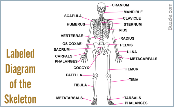
Forearm and hand bones labeled diagram labelled radius bone. Correctly label the following anatomical parts of.,label the long bone skull, clavicle, mandible, scapula, thorax, sternum, humerus, ulna, radius, carpus, phalanges (fingers), metacarpus, spine, pelvis, sacrum, femur, tibia.